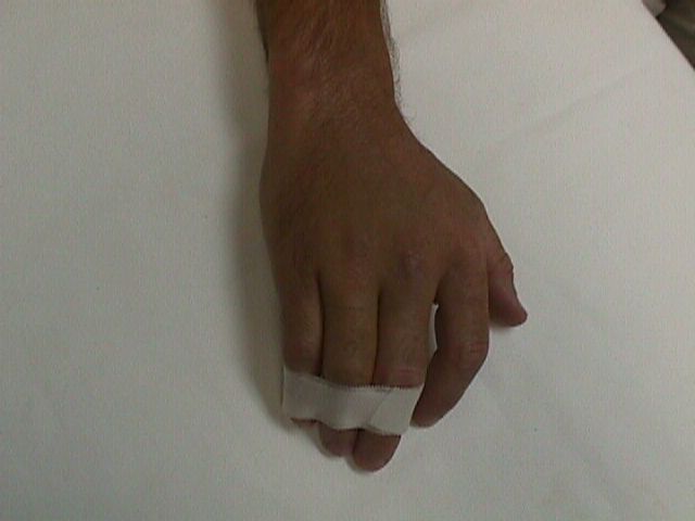
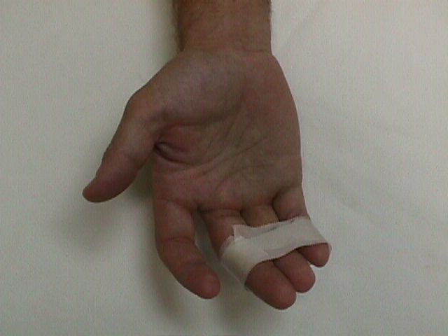
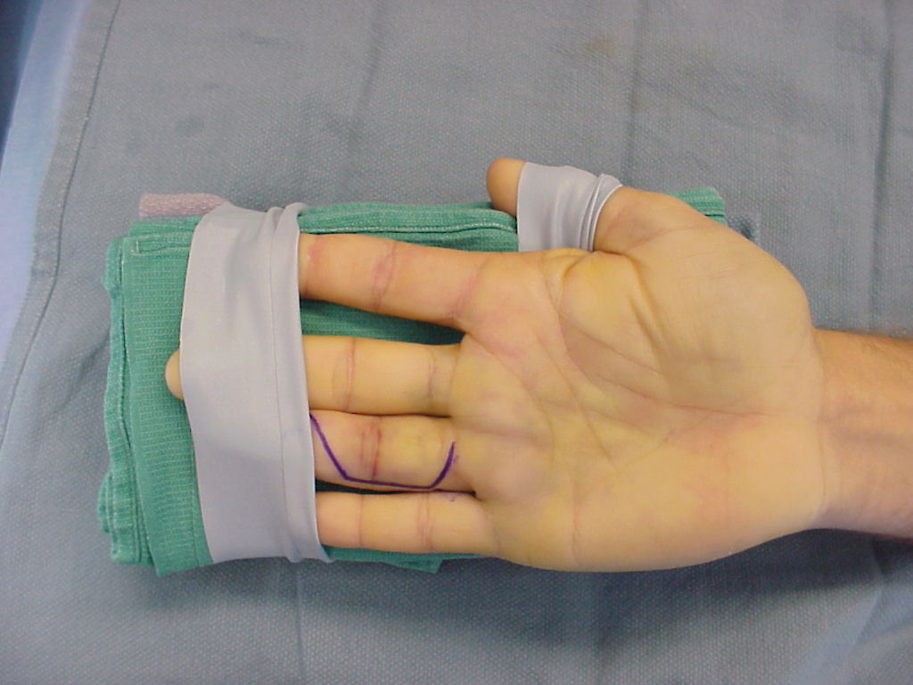
| This gentleman sustained a closed severe torsional injury of the right ring finger. |
| Click on each image for a larger picture |
 |
 |
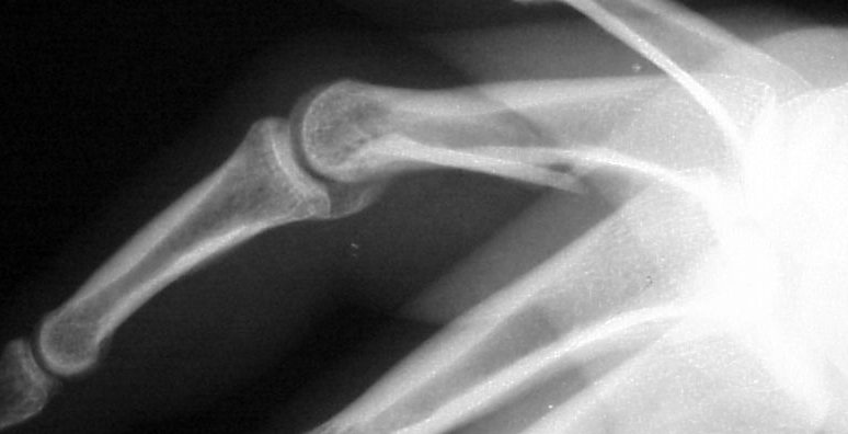
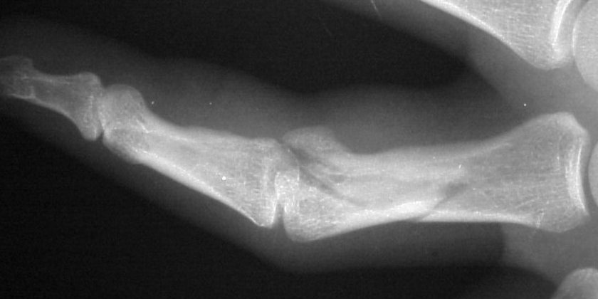
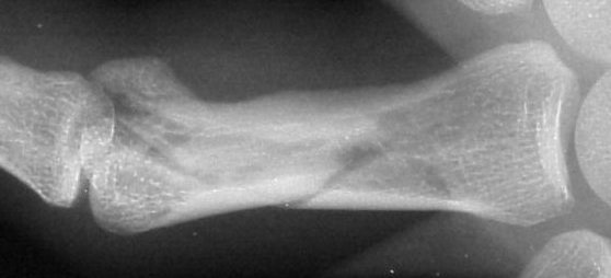
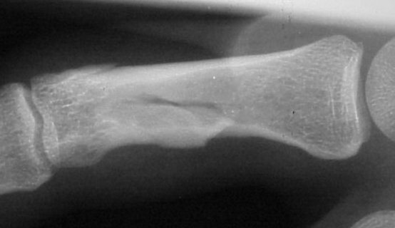
| Xrays show a complex intraarticular fracture of the proximal phalanx head. There is a displaced spiral oblique longitudinal fracture with a second oblique coronal split through the articular surface of the radial condyle. |
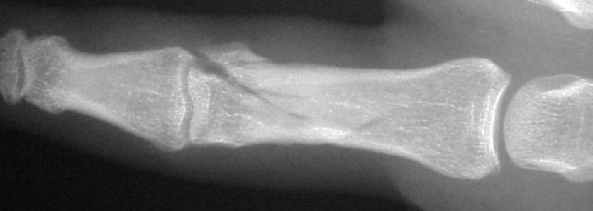 |
| Palmar displacement of a portion of one condyle is visible on the lateral view. |
 |
 |
 |
 |
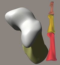
| The heads of the proximal and middle phalanges have a complex double wheel (condyle) contour,similar to that of the knee. Motion requires proper three dimensional alignent of all surfaces: |
 |
| This fracture was exposed through a volar approach similar to that used for a volar plate arthroplasty. |
 |
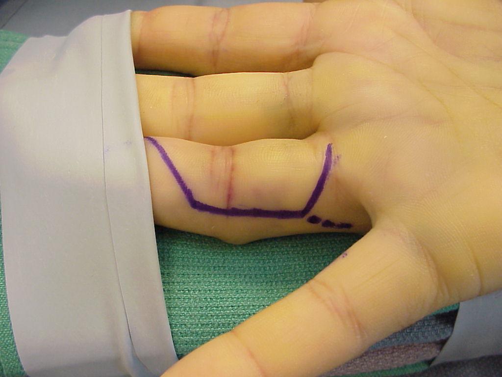
| A possible "Y" access is planned here as the dotted line. |
 |
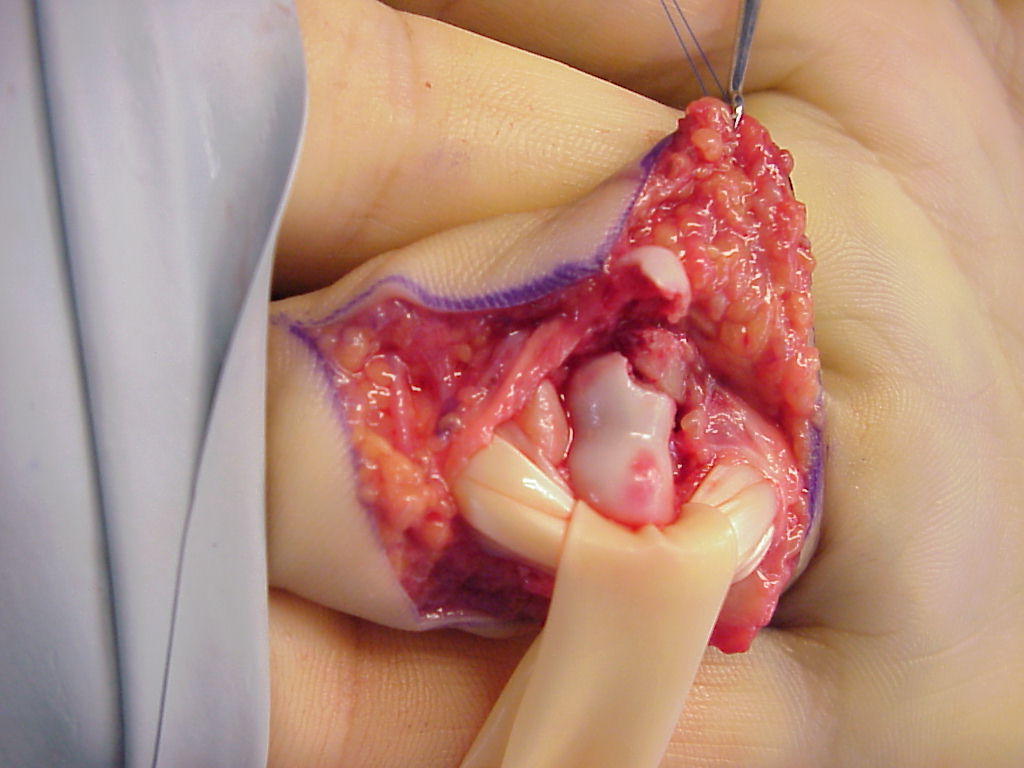
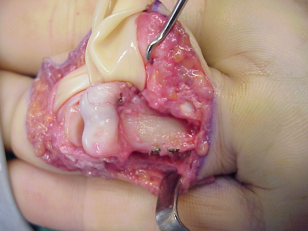
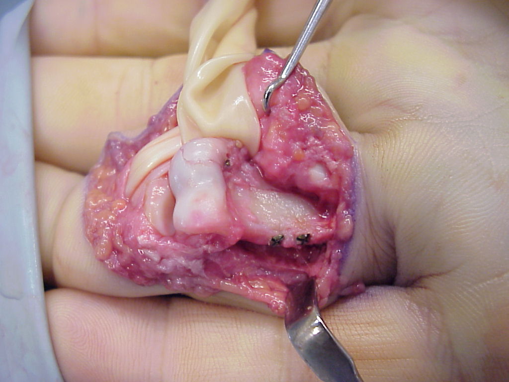
| The proximal phalanx head was in two main fragments, separated by a spike of bone from the proximal shaft. Flexor tendons are retracted here with a small Penrose drain. |
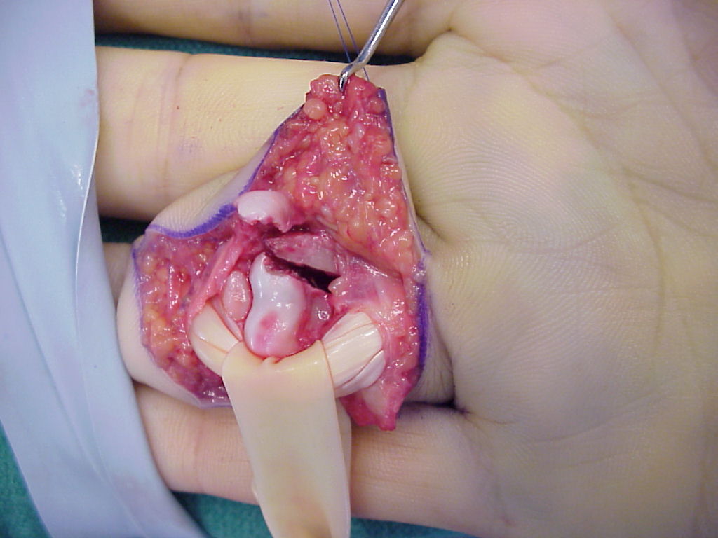 |
| Contusion of the ulnar condyle. |
 |
| The joint was reconstructed with 1.0mm screws and the shaft fixed with 1.3mm screws using the Synthes titanium modular hand tray. |
 |
 |
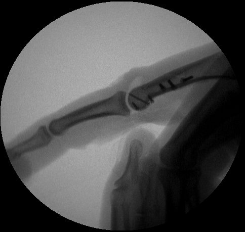
| Intraoperative fluoroscopy. |
 |
 |
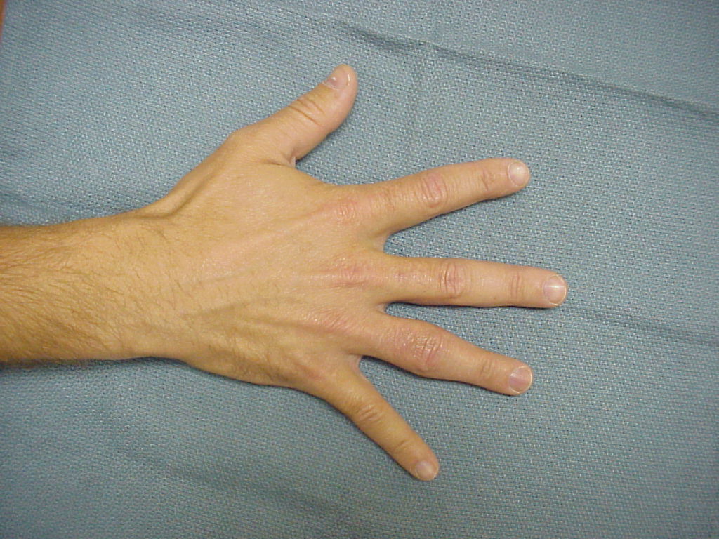
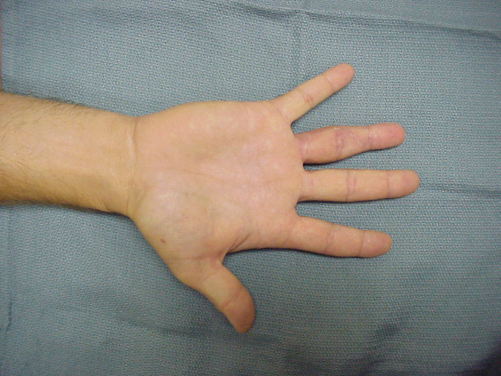
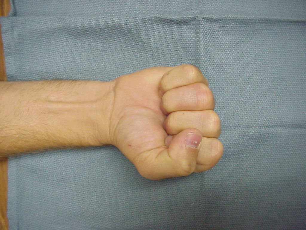
| Six months after surgery: |
 |
 |
 |
 |
 |
| Search for...
proximal phalanx head fracture bicondylar proximal phalanx fracture Synthes modular hand fixation |
Case Examples Index Page | e-Hand Home |