Clinical Examples: Fat Pad and Palmaris Brevis Flaps for Recurrent Carpal Tunnel Syndrome
| Revision surgery may be required after carpal tunnel release because of persistent or new symptoms following surgery. When postoperative problems are thought to be due to incomplete release, nerve damage during surgery, or scar tissue tethering nerve excursion, open release is indicated, often paired with an adjunct procedure. Local flaps may be used to increase nerve vascularity and reduce pain from scar tethering. Cases below show variations on palmaris brevis and hypothenar fat pad flaps in this context. Each of these patients had symptomatic relief from the procedure described. |
| Click on each image for a larger picture |
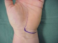
| Case 1. Palmaris brevis flap. This patient had worsening tenderness, pain and paresthesias following open carpal tunnel release done elsewhere. The old incision (dotted) was used with a proximal transverse extension. |
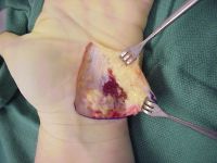
| Palmaris brevis exposed. |
| Transverse retinacular ligament released and palmaris brevis mobilized on ulnar artery perforators. |
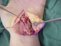
| Epineural neurolysis. |
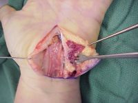
| Radial border of muscle sutured to under surface or radial leaf of transverse retinacular ligament. |
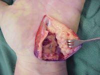
| Closure. |
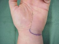
| Late result. The hypothenar skin flap was indurated for several months before resolving, one of the issues with this particular technique. |
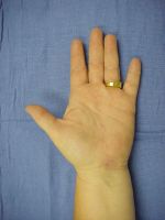
| Case 2. Combined palmaris brevis and hypothenar fat pad flap. Referred for persistent local tenderness and sudden electrical paresthesias with gripping and wrist excursion following open carpal tunnel release. Incision similar to case 1. |
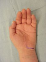
| Fat pad exposure. |
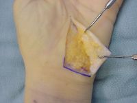
| Fat pad elevated en bloc with palmaris brevis muscle. |
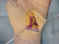
| Ulnar artery nutrient perforators. |
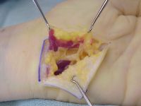
| Surface of the flap; epineural neurolysis of the median nerve. |
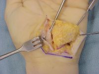
| The flap is turned over like a page in a book on an ulnar artery axis, the fat pad surface facing the nerve. The original ulnar border of the flap is sutured to the undersurface of the radial leaf of the transverse retinacular ligament. |
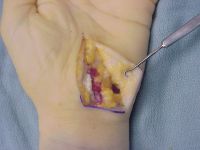
| Late result. |
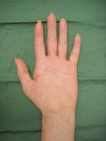
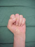
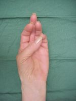
| Case 3. Hypothenar fat pad flap. This patient was referred for constant paresthesias and hypesthesia following endoscopic carpal tunnel release performed for nocturnal paresthesias. |
|
|
Two minute video summary:
|
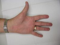
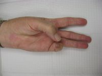
| Late result. |
|
Search
for... recurrent carpal tunnel fat pad flap palmaris brevis flap |
Case Examples Index Page | e-Hand Home |