Clinical Examples: Osteoid Osteoma
| Osteoid osteoma is a benign
painful skeletal tumor of unknown etiology which most often occurs in
the extremities. Radiographs usually show a small sclerotic bone island
within a circular radiolucent area. They may be associated with
adjacent soft tissue swelling, endosteal and cortical sclerosis.
Classically, they are painful, and the pain responds dramatically to
aspirin. These are benign, frequently do not progress, and pose little
health risk. Excision is indicated for inadequate pain control or
questionable diagnosis. Excision margins may be difficult to estimate
because of associated adjacent sclerosis. These tumors have high rates
of bone turnover, allowing the use of metabolic bone markers such as
tetracycline or technetium, as demonstrated in these cases. |
| Click on each image for a larger picture |
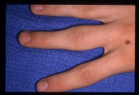
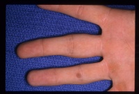
| Case 1. This 18 year old man
presented with a two year history of pain and swelling of the distal
aspect of his proximal phalanx. |
| Plain films showed
sclerosis within a radiolucent area and adjacent cortical/endosteal
sclerosis. |


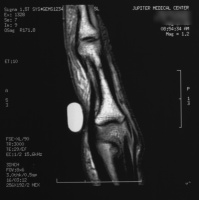
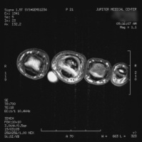
| MRI confirmed a cortical
nidus. |
\
| The tumor was removed with
a burr. |
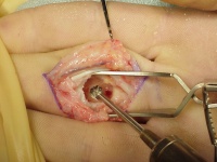
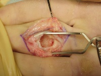
| Despite extensive
resection, over three quarters of the cortical circumference remained,
and structural reinforcement was not necessary. |
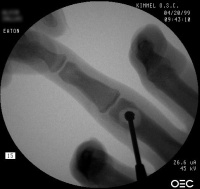
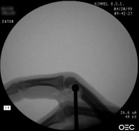
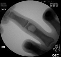
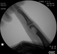
| The defect was packed with
cancellous bone harvested from the dorsal distal radius. |

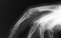
| Case 2. (Case of Charles Melone MD) This patient presented with a pathologic scaphoid fracture associated with an osteoid osteoma. |
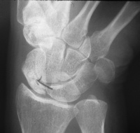
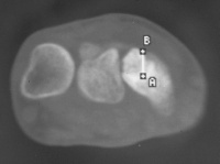
| This was managed with
scaphoid excision and 4 corner fusion. The patient was given oral
tetracycline preoperatively to aid in tumor marking. This view is
of the distal scaphoid, looking at the fracture line. |
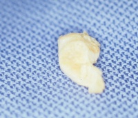
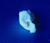
| The same view in
ultraviolet light shows light yellow fluorescence of the central nidus. |
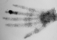
| Case 3. This 26 year old
woman presented with recurrent pain and swelling two years following
excision of an osteoid osteoma of the ring finger middle phalanx head.
Bone scan shows intense activity in this area, consistent with either
persistence or recurrence of osteoid osteoma. |
| The patient was given
perioperative technetium and tumor resection was guided using a
hand held technetium probe. |
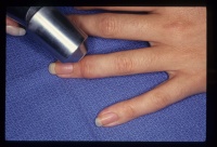
| The tumor was resected
using a dorsal tendon splitting exposure. |
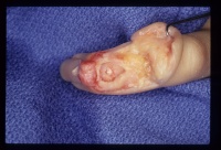
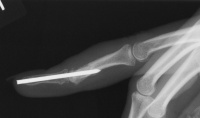
| Debridement was continued
until normal counts were demonstrated . This left a thin
cortical shell which was reconstructed with a corticocancellous graft
from the distal radius. |


| Search
for... osteoid osteoma hand osteoid osteoma technetium |
Case Examples Index Page | e-Hand home |