Clinical Example: Nail complex reconstruction with nail bed grafts and reverse partially deepithelialized dorsal cross finger flaps for eponychial reconstruction
| Fingernail anatomy is
complicated. Even more complicated is the task of reconstructing
components of this area. One of the trickier issues is the eponychial
fold, which has skin on both sides. One technique for replacing this
area is a reversed deepithelialized dorsal cross finger flap, which is
demonstrated with this case. |
| Click on each image for a larger picture |
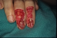
| This 40 year old man on
insulin for diabetes sustained loss of the dorsal soft tissues of his
dominant index and middle fingers in a rotating blade injury. The
central nail beds and central eponychial skin were partially
lost, as was the dorsal DIP joint capsule and terminal tendon of the
middle finger. |
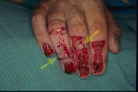
| The nail beds were
reconstructed with nail bed grafts from the adjacent fingers. the
middle finger terminal tendon was reconstructed with a slip of lateral
band used as a graft inserted distally with a bone anchor. Dorsal flaps
were elevated and partially deepithelialized, leaving distal areas
undisturbed to reconstruct the undersurface of the eponychial skin fold
(yellow arrows). |
| Donor sites and flaps were
closed with a full thickness skin graft from the medial proximal
forearm. Nail beds were dressed with Gelfilm. |
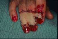
| Two weeks postop,
immediately prior to flap division. |
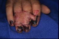
| Two weeks after flap
division. |
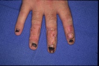
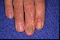
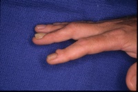
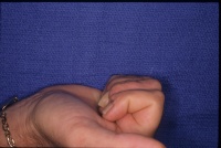
| One year postop, after debulking flap revisions. |
|
Search for... reverse dorsal cross finger flap eponychium reconstruction |
Case Examples Index Page | e-Hand home |