Posting | Hardware | Software | Formats | Optimizing files | Optimizing quality
Posting 
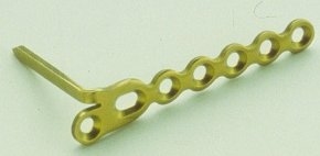
an image in a mailing is just a matter of including the web site address of the file in the mailing. For email, it's best to reference the location of the image, which is a snap once you have the image archived on a web site: just type in the web address of the image, like this: HTTP://www.eatonhand.com/jpg/jpg01018.jpg and in the current email programs, the conversion to a link is automatic - just type in the address and the program makes the link. That way you can put in a number of pictures without clogging (and possibly crashing) the recipient's mailbox. Getting the actual image up onto the  web site is a different matter. You can only do this from a web site which is up and running.

If you don't have your own web site, you have two options:
1. Start your own personal web page - you can do this for free through HTTP://www.tripod.com , HTTP://www.geocities.com and other hosting services for free, HTTP://www.aol.com.com and other commercial hosting services for a fee. In the process of setting up your page, you will be given instructions on how to post images.
2. Make arrangements with someone you know or a hospital web site to put images on their web site in your own separate directory. Whoever runs the web site should be able to either do it or walk you through it.
3. Free server space for images is available through several sources, such as http://www.photopoint.com/

Posting | Hardware | Software | Formats | Optimizing files | Optimizing quality
Hardware: However, before you can post images, you have to have image files. Slides or X rays need to be scanned into the proper file format for web use - jpg format. Most companies charge a fair bit for doing this, which is why I do my own, but if I were only doing this occasionally, I would have someone else do it - it otherwise isn't worth the trouble, getting a scanner, tweaking the picture, etc. Your hospital may be able to accommodate you - most VA hospital medical illustration departments eat this stuff for breakfast.

What I use:
Clinical (color) pictures
Digital Cameras
Sony Mavica - (original version), which has too poor resolution for any detail work, but saves to a floppy.
Casio QV-10 + Snappy video to parallel port capture. Too poor resolution for any detail work, but absolutely least expensive system.
Sony Mavica MVC-FD81, which has a resolution of 1024 X 768 pixels (.78 megapixels) and will also take up to 6 second MPEG movies, saves to a floppy. This has replaced slides as my primary photo tool - It is satisfactory for both clinical and Xray use.
Nikon Coolpix 990, which has higher resolution (3.34 megapixels) and better focusing algorithms than the Mavica, but a poorly designed flash which casts strong shadows on extreme closeup shots, making it unsuitable for intraoperative macro shots. Disappointing.

Scanning Slides

Slides: I use an HP Photo Smart Slide scanner. This produces too much artifact on slides of X rays, but is very easy to use and very slick for clinical pictures.

What to do for Xrays
Taking slides of X rays: I use Fujichrome ASA 100 (cheap) and manually set my shutter speed at a 45th of a second or longer to avoid taking a picture of the black line which travels down the fluorescent bulb at 50 -60 cycles per second. I then set the aperture on automatic. When shooting close ups with this setup, pay attention to the aperture your camera chooses, and increase the exposure time as needed to keep the aperture within your camera's range - e.g. the number should be greater than 2.8. Use a copy stand for exposure times longer than 1/10 second.
Scanning slides of X rays: I use a Microtek ScanMaker 35t plus. This unit is slow and a bit difficult to adjust, but very powerful, and can provide extreme magnifications - much better resolution than the HP unit above.
Scanning Regular X rays: Flatbed scanner with transparency adapter: I have tried the HP Flatbed scanner, but surprisingly, this can produce artifact and crashes the computer when the Xray film is larger than and overlaps the scanning. 
The transparency attachment is always a special order option for flatbed scanners, and is not stocked in the stores. I have been told that shining a bright light on the Xray while scanning can substitute for a transparency adapter, but haven't tried it myself. (I have used the same trick with limited success making photocopies of X rays).

My advice: if you're not sure how much you will be using it, outsource to some camera or copy shop as a test drive. If you really want to do it yourself, look into flatbed scanners with transparency adapters. If you are using a PC, the best setup is to use a scanner with a SCSI card, which has to be installed into the computer. It will save time in the long run. Parallel port scanners work well and are inexpensive, but are slow - if it takes 5 minutes per scan and you have 10 X rays and 3 of them have to be re scanned... do the math. If you're on a Mac, get a scanner now!

Posting | Hardware | Software | Formats | Optimizing files | Optimizing quality
Software used on this site is detailed here
Posting | Hardware | Software | Formats | Optimizing files | Optimizing quality
Image Formats
Images must be in either GIF or jpg format to work on web pages.
GIF format is best for high contrast line drawing or text images.
jpg format is best for photographic type images.
Using jpg for line drawings results in an image which is either more blurry or much larger than its GIF counterpart:
GIF Image, file size 11K. This high contrast image is best suited for GIF format.
Best jpg version, file size 44 kb (no compression) - not quite as sharp, and the uncompressed image size is four times that of the GIF version.
jpg version, compressed to the same size as the GIF version (file size 11 Kb), which required a compression factor of 60. Compression artifact is most noticeable in high contrast line art - a poor choice for jpg. 

Using GIF for photographic type images results in files which are larger and slower to download than jpg files of the same picture:
jpg format, good quality (compression factor 6), file size 13K. This variable contrast photo style image is best suited for jpg format.
GIF format: file size 25K. Same quality image, but the file size is twice as big, and so twice as long to download - photo images are usually the wrong use for GIF format for this reason. Additionally, GIF format limits the total number of colors to 256, which can result in a "poster" or "contour map" appearance, losing in subtle color gradients.
 

Posting | Hardware | Software | Formats | Optimizing files | Optimizing quality
Optimizing Image Size for Web Use:
Scanning resolution
Slides: depending on the menu options allowed, and depending on how much I'm zooming in on the slide, I scan slides at resolutions of
400 dpi (usual) to 800 dpi (max), or
600 pixels wide (usual) to 900 pixels wide
Image Dimensions
Physical size: I try to keep the image smaller than 400 pixels tall and 550 pixels wide, so that the entire image will fit on most monitors without needing to scroll.
X rays: Most hand, wrist and elbow X rays will fit on the computer screen life size. Set the flatbed scanner to a 100% image size for most, 200% for fingers, 50% for forearms.

File Size
I try to keep the file size under 50 kb so that loading isn't slow. There are a few trick to doing this - in this sequence:
Crop the image as much as possible without losing landmarks for orientation.
Set X rays to monochrome or grey scale.
Set Xray compression ratio to 15, other images to 25.
Last resort, increase jpg compression ratio. *Some programs and cameras set the jpg compression ratio at maximum (uncompressed) by default, which results in the largest possible file size. Most clinical pictures can be compressed by adjusting the jpg compression ratio to reduce file size 50 to 75% without noticeable loss of image quality - . , which 

Posting | Hardware | Software | Formats | Optimizing files | Optimizing quality
Optimizing Image Quality for Web use:

The scans are usually not perfect right out of the scanner or camera. Here are some tweaks to optimize scanned images for web display. I use Paint Shop Pro, but all scanners and most digital cameras come with image editing software. My current approach is to modify in this order:

  1. Open the image in an editing program.
  2. Save as a new file so that the original is maintained as an archive.
  3. Crop, but leave landmarks for orientation.
  4. Resize to monitor fit: e.g. smaller than 400 pixels tall and 550 pixels wide 
  5. Resizing usually blurs the image. Correct this differently, depending on the image:

  6. either
    Sharpen clinical images or plain Xrays.
    or
    Edge enhance for CT or MRI images.
  7. Grey scale convert Xray, CT, MRI - this reduces file size and makes the image look better.
I know that some of this may sound like gobbledegook, but those are the kinds of editing controls paint programs have. The exact command name and options will be different for other programs.

Note: In general, images obtained from low end digital cameras (image size 640 X 480 pixels or less) are so muddied by compression that they can neither be sharpened nor made more clear with any photo editing software.

I don't have all the bugs worked out myself - variations in X ray exposure seem to make a big difference in the quality of the scanned images - for example, a coned down finger view may give a great scan, but the greater exposure the tech uses for a wrist PA may make it impossible to obtain a comparable finger pic from an X ray of the whole hand - even though it is the same finger and the same Xray magnification.

Where is that picture?

Keep a database of which patients have which slides. I use ACDSee for this, which lets me keep a file of descriptions of each image file. I make a point of writing the image file number right on the slide for later reference.

So - before you spend serious bucks, sit down and figure out what you want to keep track of for the next 20 years and set up your database - for me, it's simple, plain things such as:
 

0 - patient's name/unique identifier.
1 - Diagnoses.
2 - Operations.
3 - Misc. notes about special findings which aren't easily categorized, but which will drive you crazy when you try to find them in 10 years (e.g. who was that patient who had a capitohamate coalition in the exact shape of Pamela Anderson's left breast in Baywatch episode # 63?).
4 - Xray findings.
5 - Notes about whether the file is complete/closed (e.g. "need a late Xray showing final growth plate remodeling", or "need final appearance of the reconstruction" or "Nice - a complete set" - that is what sets the reports of the masters apart from those of the amateurs - the late documentation).
6 - viewable digitized images.

Then, a way to enter in the info - at some point, sit down with your Dictaphone and your old slides, dictate these items (database fields), and have someone enter them into your database, transcribing them from your dictation - that will get you caught up to the present, then either continue with that or design a paper or electronic form to fill out as you go, to be entered into your database to keep current for the rest of your practice, and -easy - the job is on autopilot!

None of this stuff is hard, but like most computer things, it takes many, many little steps - unfortunately, more than I can just run through here, because the exact steps will vary entirely on your exact scanner / software setup. There is an unavoidable portion of the learning curve you just have to either learn it yourself (there are many books on setting up a web site, etc.), have a local geek teach you, or out source it. I hope that this helps some.

Charlie Eaton