| Dorsal fracture dislocations of the proximal interphalangeal joint are a common and difficult injury. The seriousness of the problem is often underestimated by the patient, leading to delay in treatment. Even when treated early, these fractures are difficult because the joint surface is crushed into small pieces. Treatment options include - among others - fusion, dynamic traction, ORIF with bone graft, volar plate arthroplasty, and joint reconstruction using a hamate osteochondral graft matched to the shape of the base of the middle phalanx, which was used in this case. There is no single solution which works well for all patients. |
| Click on each image for a larger picture |
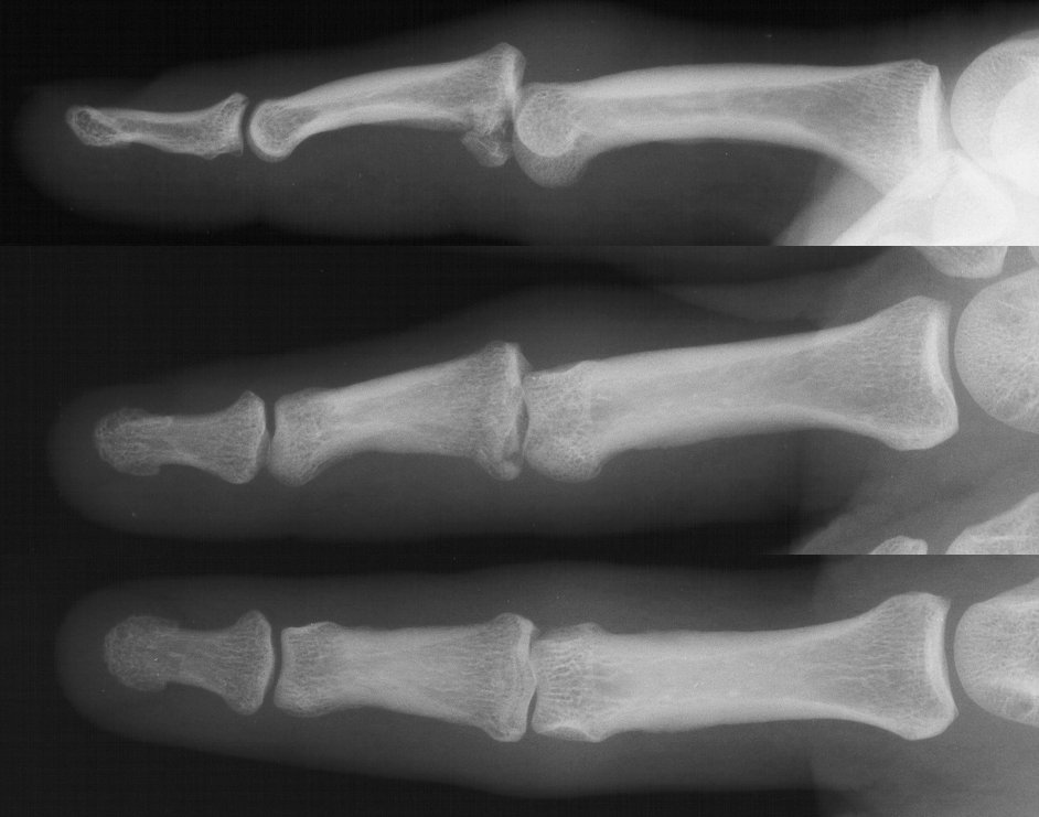
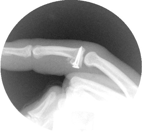
| This gentleman sustained a comminuted proximal interphalangeal fracture dislocation. |
 |
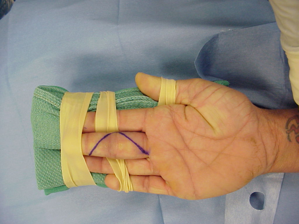
| The incision. The tip of the flap has unpredictable healing whether it is planned as a "V" here or as a longer longitudinal midaxial incision. |
 |
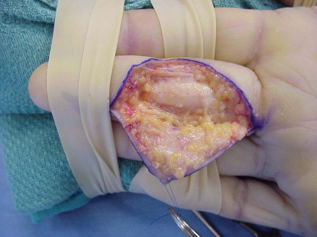
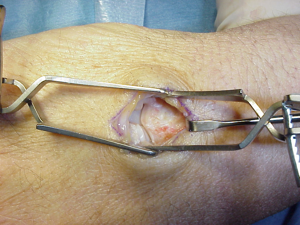
| The flexor tendon sheath and neurovascular bundles are exposed. A window is oened in the sheath to excise the collateral ligaments and detach the distal insertion of the volar plate. |
 |
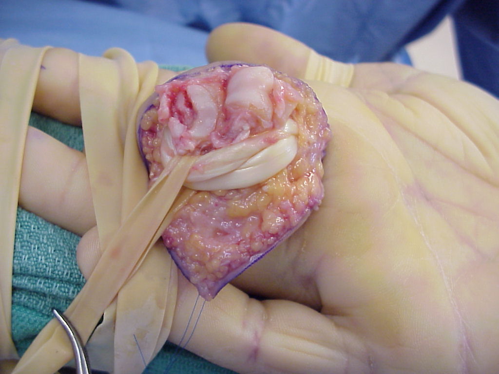
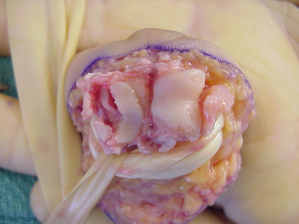
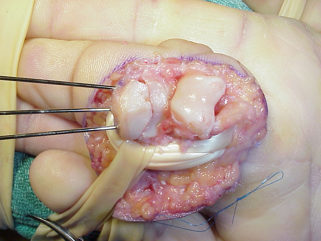
| This allows the joint to be hyperextended 180°, fully exposing both joint surfaces. The flexor tendons are retracted ulnarly with a penrose drain. |
 |
| The palmar base of the middle phalanx is in at least three thin fragments. The palmar ulnar condyle of the proximal phalanx head has irregular sections of cartilage loss. |
 |
| The dorsal hamate carpometacarpal area is used as a donor site for joint surface reconstruction. It has a "gull wing" biconcave contour which usually matches that of the missing finger joint surface. |
 |
| Here, the graft has been harvested and sculpted to fit the remaining joint. Three 0.035" K wires are in place for provisional fixation. |
 |
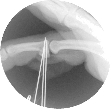
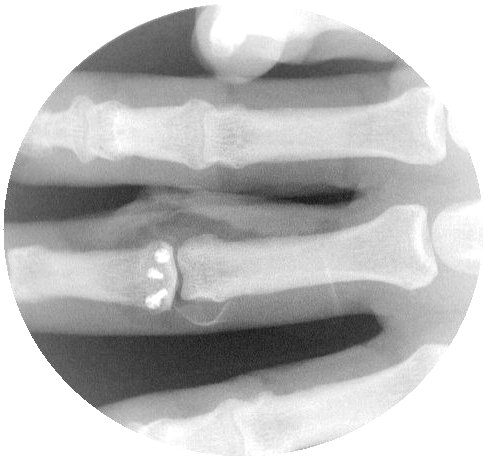
| Intraoperative fluoroscopy confirms proper realignment of the joint with the bone graft in place. |
 |
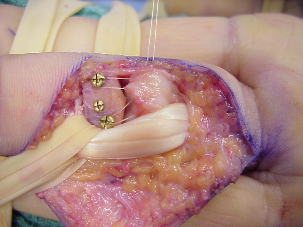
| The pins are replaced with screws, one at a time. Here, two 1.3mm and one 1.0mm screws were used. |
 |
 |
| The last step before repairing the flexor tendon sheath is reattaching the volar plate. One method is to slightly back out the lateral screws, loop a suture from each corner of the volar plate around them, tighten and tie the suture, then advance the screws back in. The third screw provides extra security during this step. |
 |
|
here and here |
| Search for...
hamate osteochondral graft proximal interphalangeal fracture dislocation |
Case Examples Index Page | e-Hand Home |