Clinical Example: Reduction of Locked Metacarpophalangeal Joint
| Locked metacarpophalangeal
joints are uncommon. The most common anatomic pathology is entanglement
of a collateral ligament on an osteophyte at the base of the proximal
phalanx. Most often this occurs spontaneously during use. In most
instances, closed reduction is possible. Open reduction may be necessary |
| Click on each image for a larger picture |
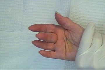
| This 75 year old healthy
woman had a sudden event reaching into a drawer two days ago. Since
then, she could not straighten her middle MCP. |


| Standard AP Xrays were
unremarkable. |

| However, an AP view with
the proximal phalanx flat against the plate perpendicular to the beam
shows prominent proximal phalanx osteophytes, one of which has caught
one of the collateral ligaments. |

| Closed manipulation was
performed with a median nerve block, rotating the proximal phalanx
through pronation and supination while applying longitudinal
distraction in the direction of the deformity. Click on the image below
for a video of the reduction. |
| Search
for... locked metacarpophalangeal joint |
Case Examples Index Page | e-Hand home |