| Distal radius fractures are most likely to give long-term symptoms when malunion involves intra-articular incongruity, shortening, or residual dorsal angulation. Frequently, shortening results in malalignment of the distal radial ulnar joint, and may result in ulnolunate impaction syndrome. If joint surfaces are preserved, these last two problems may be corrected surgically with either corrective osteotomy of the distal radius or ulnar osteotomy. Ulnar osteotomy may be performed with corrective angulation, or as in this case, with simple shortening. Chronic changes in the distal radial ulnar joint may prevent reduction of the distal radio ulnar joint by traction produced by shortening ulnar osteotomy. This case demonstrates a technique in which ulnar osteotomy is combined with complete mobilization of the distal ulna to allow release and synovectomy of the distal radial ulnar joint from a proximal access. |
| Click on each image for a larger picture |
| Preoperative radiographs demonstrate radial shortening and relative ulnar lengthening and ulnar carpal abutment. |
 |
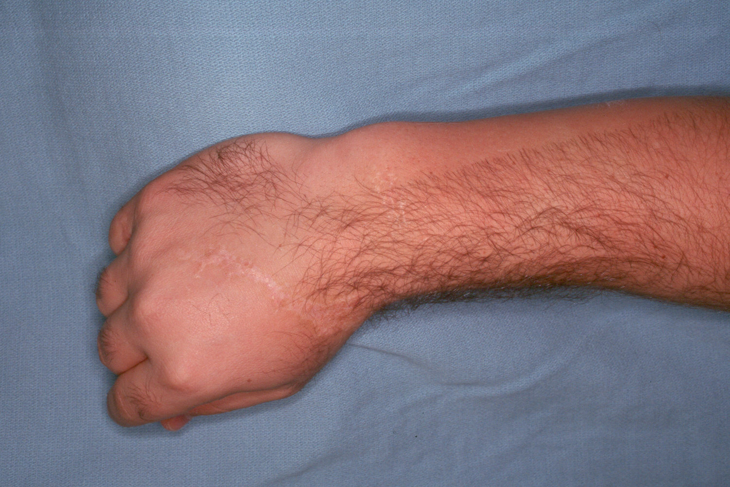
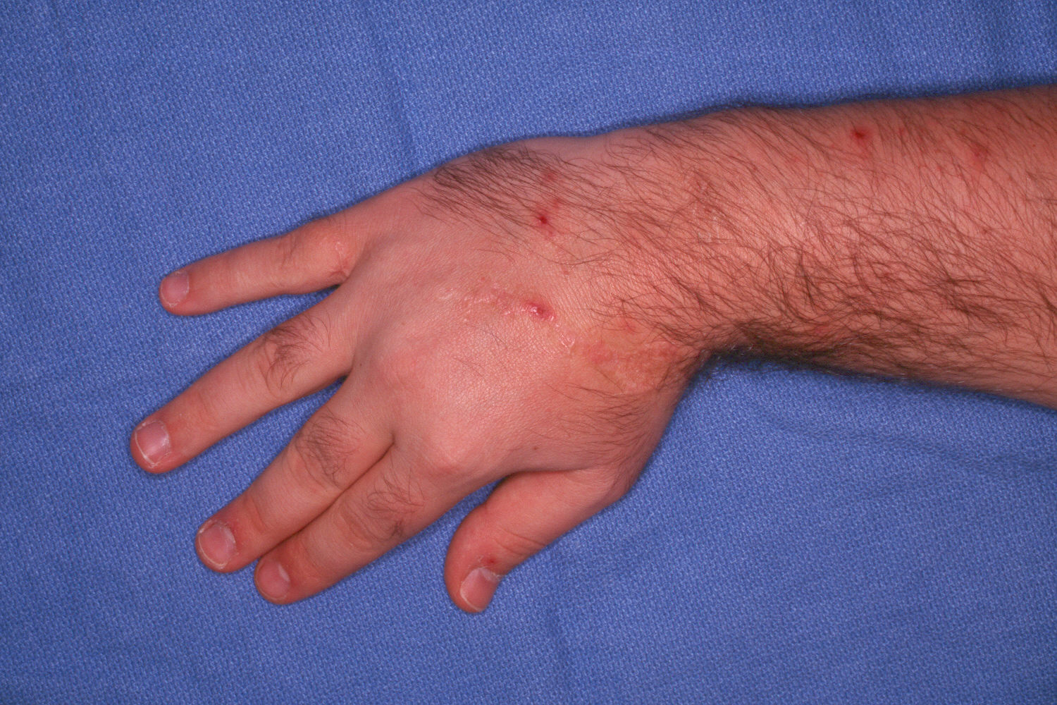
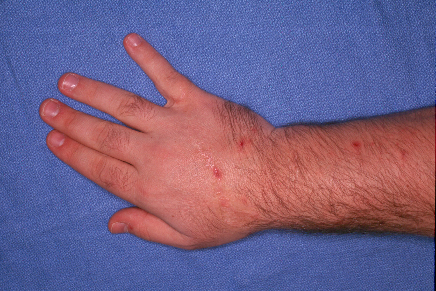
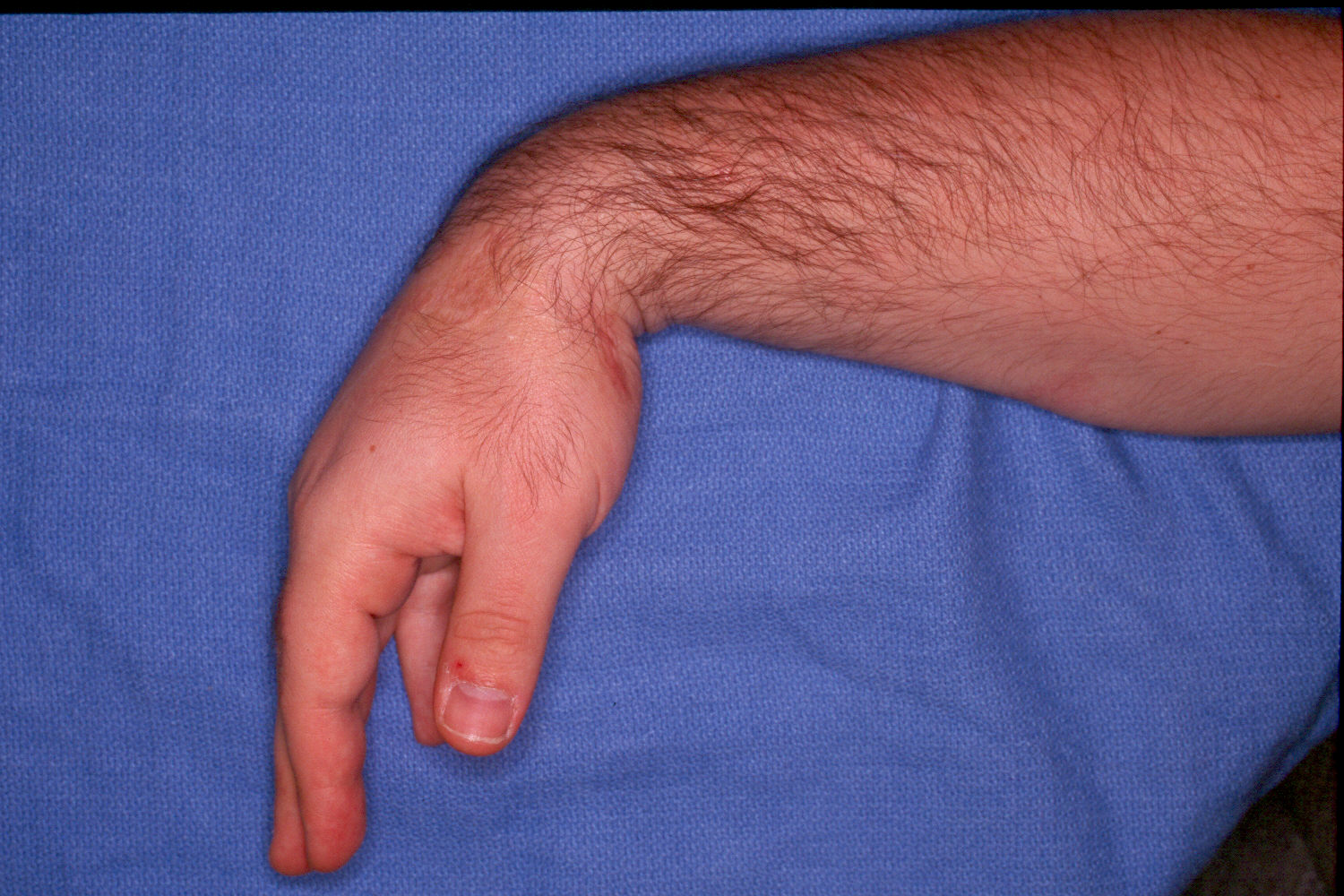
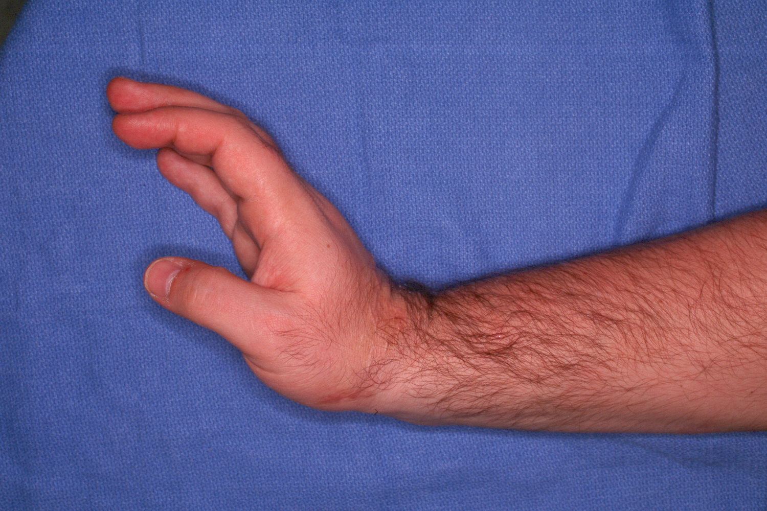
| Ulnar head prominence is obvious, associated with a fixed radial deviation of the wrist. |
 |
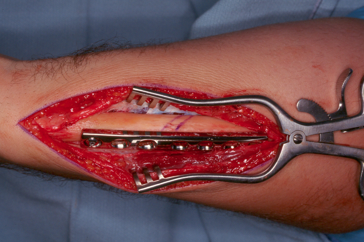
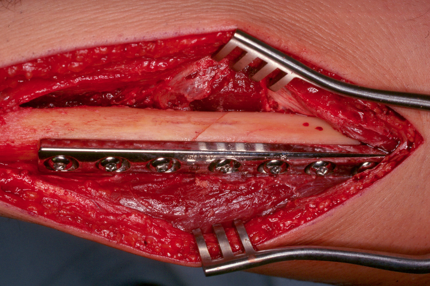
| The ulna is approached through its subcutaneous border. Provisional fixation of the plate to the distal segment is used to plan the osteotomy, which is marked here. |
 |
| An oblique section of ulna is removed with a saw. This may be done free hand by making a partial cut with a saw blade, then leaving an unattached blade in the first osteotomy path to provide a visual aid for a second parallel cut. |
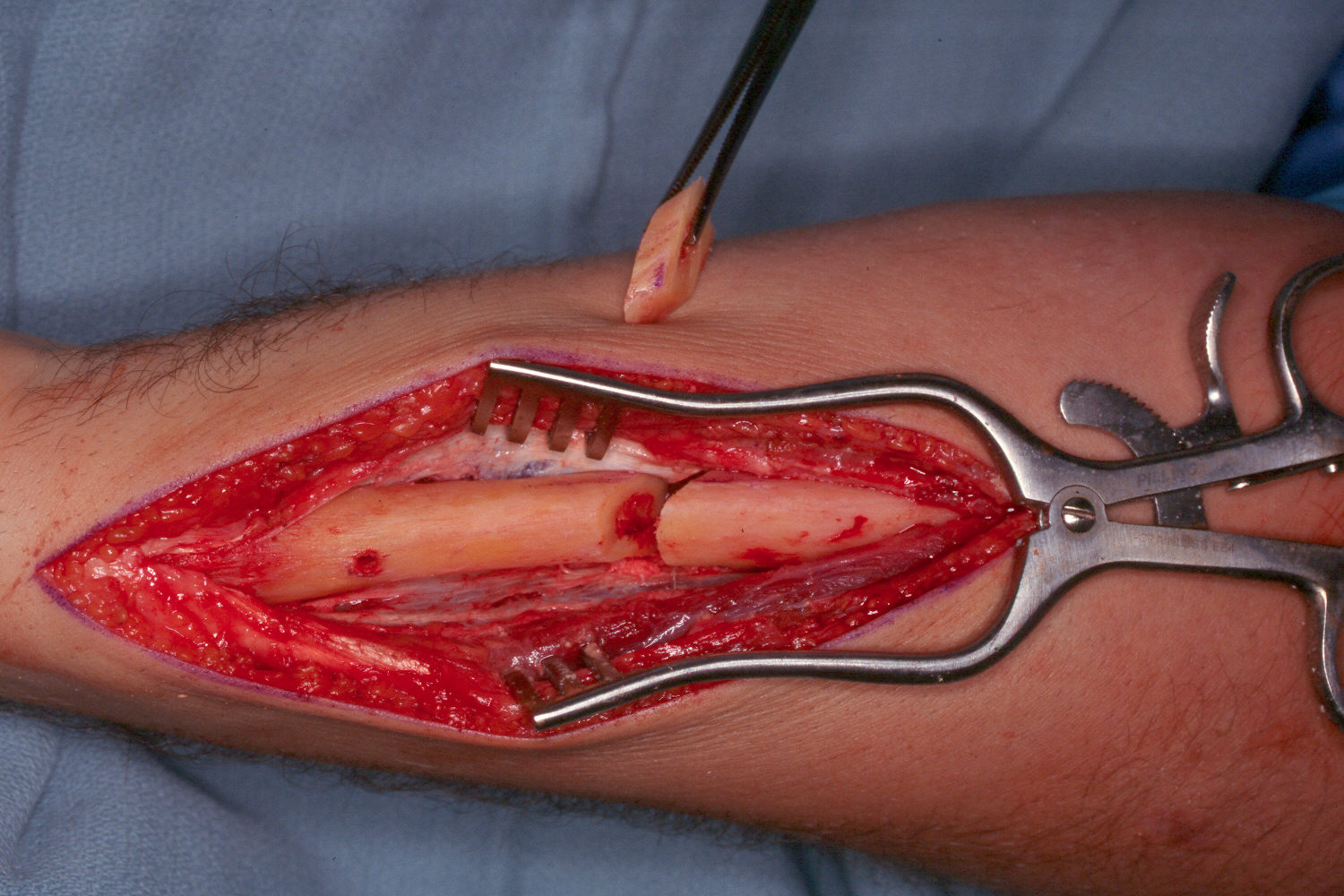 |
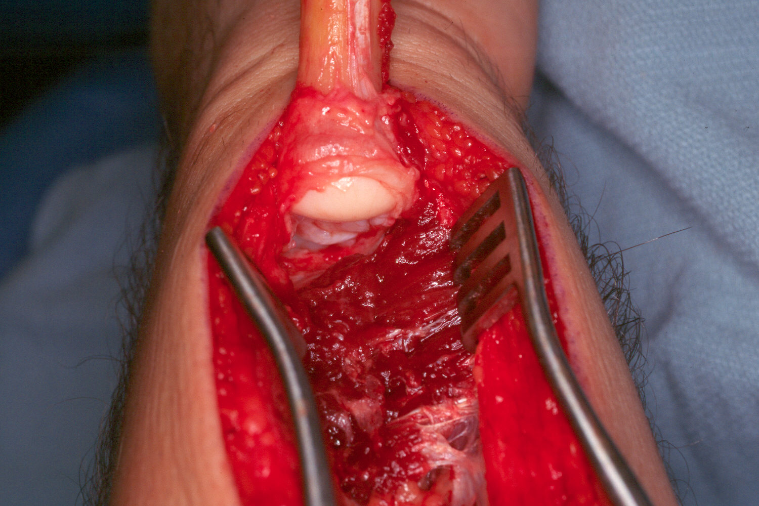
| After osteotomy, the distal ulna is freed circumferentially out through the distal radioulnar joint, leaving ulnar styloid attachments. |
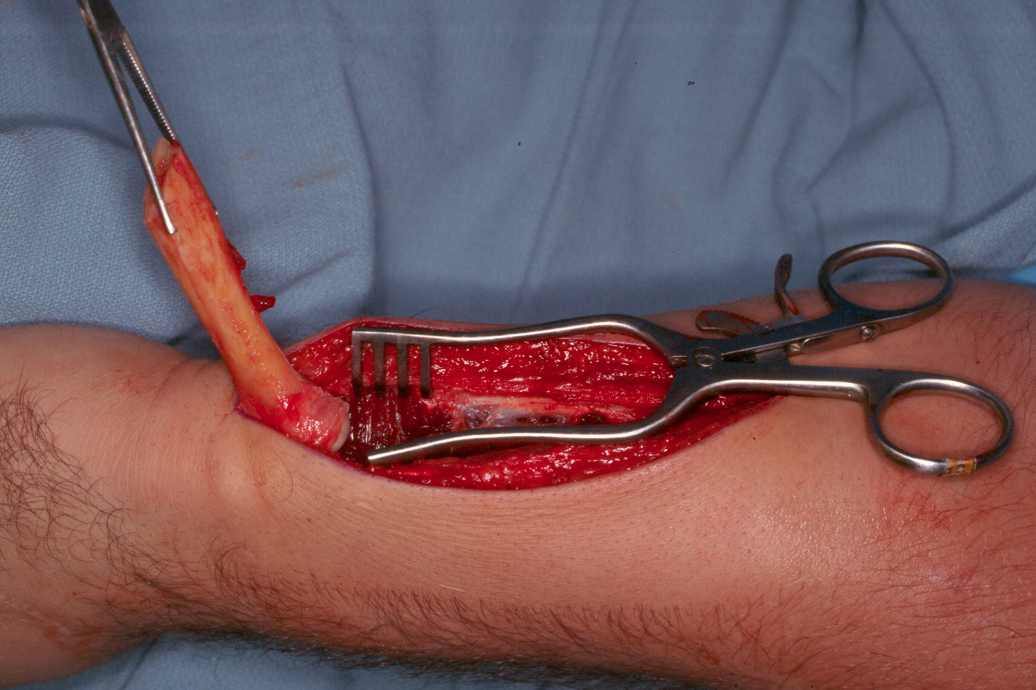 |
| The distal radioulnar joint and ulnar head can be accessed from below. |
 |
| A longitudinal saw cut partially into the cortex prior to ulnar osteotomy provides an additional aid in realignment for the final fixation. |
 |
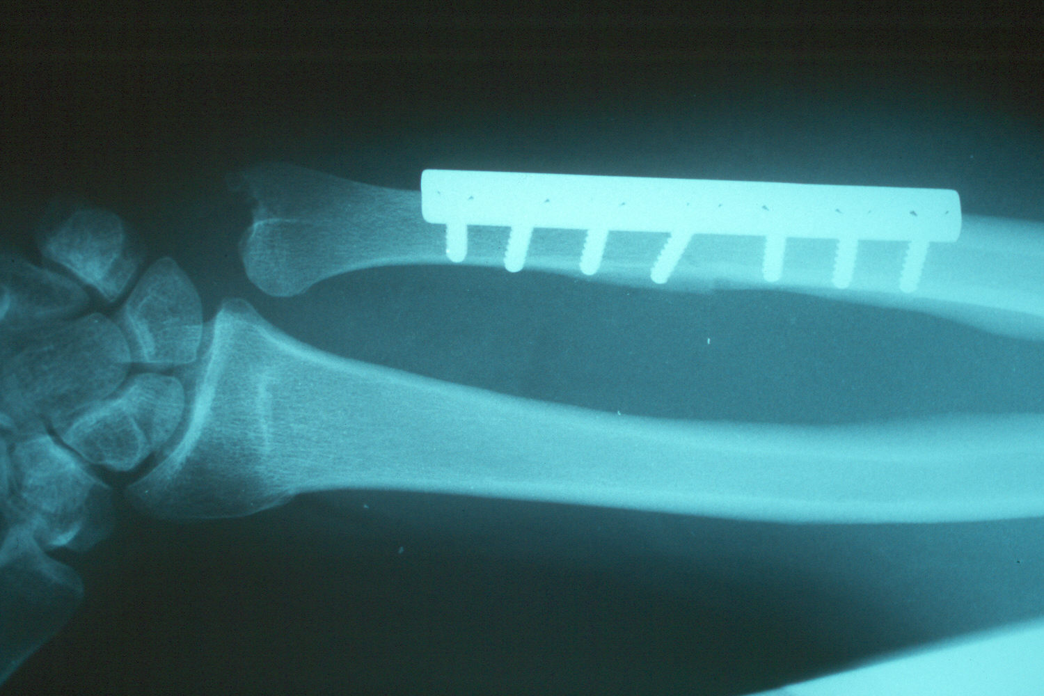
| Late result: proper ulnar alignment restored. |
 |
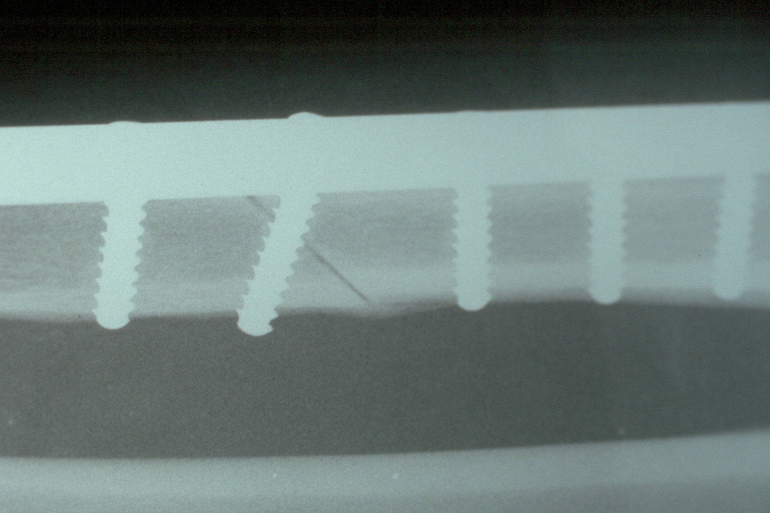
| The osteotomy and plate position are planned using a seven hole plate with a compression screw positioned obliquely across the osteotomy cut. |
 |
| Late result - healed. |
 |
| Late range of motion. |
 |
 |
 |
 |
| Search for...
ulnar shortening osteotomy distal radius malunion |
Case Examples Index Page | e-Hand Home |