| Glomus tumors are small,
painful, tender,
and interesting to hand surgeons because they are just uncommon enough
that the patient often presents with a history of having seen several
physicians,
without a diagnosis, and the hand surgeon can not only set the record
straight
that the problem is both real and curable, but also provide the
definitive
cure. Subungual glomus tumors are exquisitely tender and painful with
cold
exposure. Glomus tumors may also arise in the fingertip pulp, where
they
are usually less painful and tender. More cases are shown here. |
| Click on each image for a larger picture |
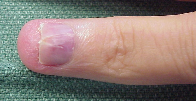
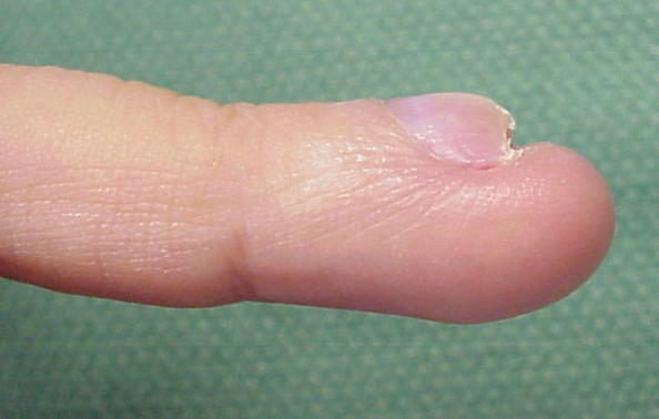
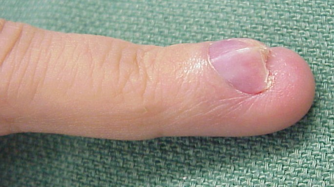
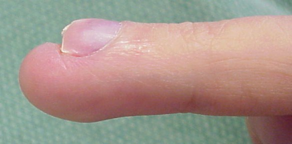
| This is the finger of a young woman who had been bothered by a painful fingertip and fingernail, accompanied by a progressive fingernail contour deformity and a pinkish discoloration of the entire proximal nail bed. |
 |
 |
 |
 |
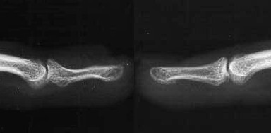
| Plain films confirmed a concave contour deformity of the dorsal aspect of the distal phalanx, shown here compared to the opposite, normal finger film: |
 |
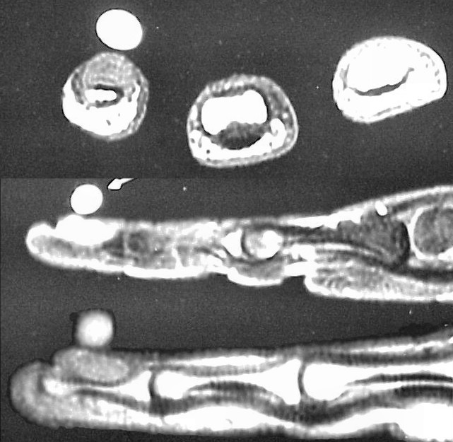
| MRI demonstrated a dorsal subungual contrast enhancing mass. |
 |
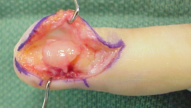
| The area was explored through a midlateral incision, exposing a large subungual tumor: |
 |
| The entire dorsum of the distal phalanx can easily be exposed through this approach: |
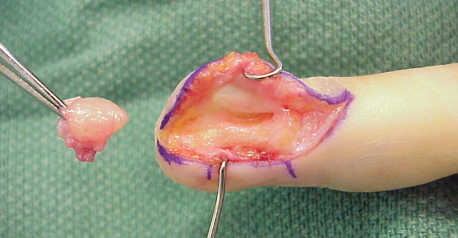 |
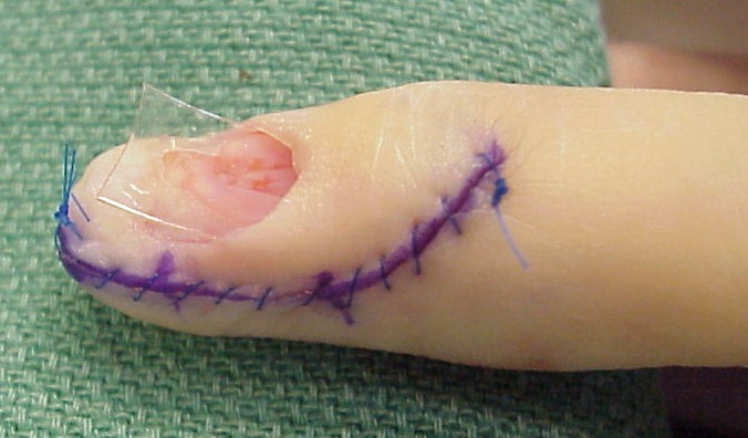
| Incision, tumor excision, nail plate removal, wound closure and gelatin sheet (Gelfilm®) placed as an absorbable nailbed spacer: |
 |
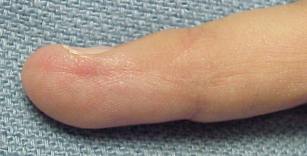
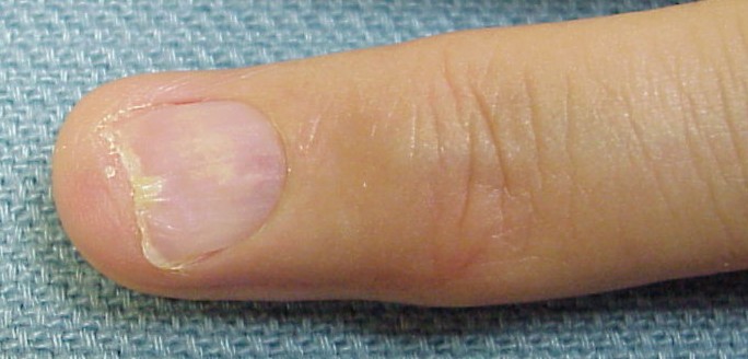
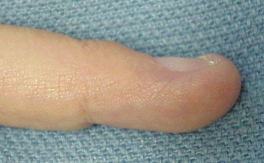
| Pathological examination of the surgical specimen confirmed a glomus tumor. These images were taken three months postop, no pain, nail plate not yet fully regrown, but growing normally: |
 |
 |
 |
| Search
for...
subungual glomus tumor glomus tumor |
Case Examples Index Page | e-Hand home |