Clinical Example: Intraarticular Distal Radius Malunion
| Because of the biplanar
angulation and curvature of the distal radius, the alignment of
intraarticular distal radius fractures may be difficult to estimate by
plain radiographs. |
| Click on each image for a larger picture |
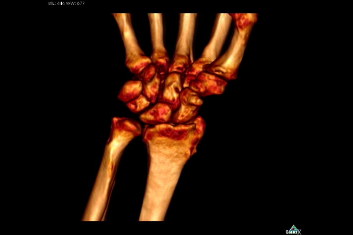
| This patient presented with
persistent wrist pain, swelling and stiffness one year out from closed
reduction and external fixation of a n intraarticular distal radius
fracture. |


| No prior films were
available. Plain films suggested a depressed section of the radial edge
of the lunate fossa and loss of dorsal tilt. |


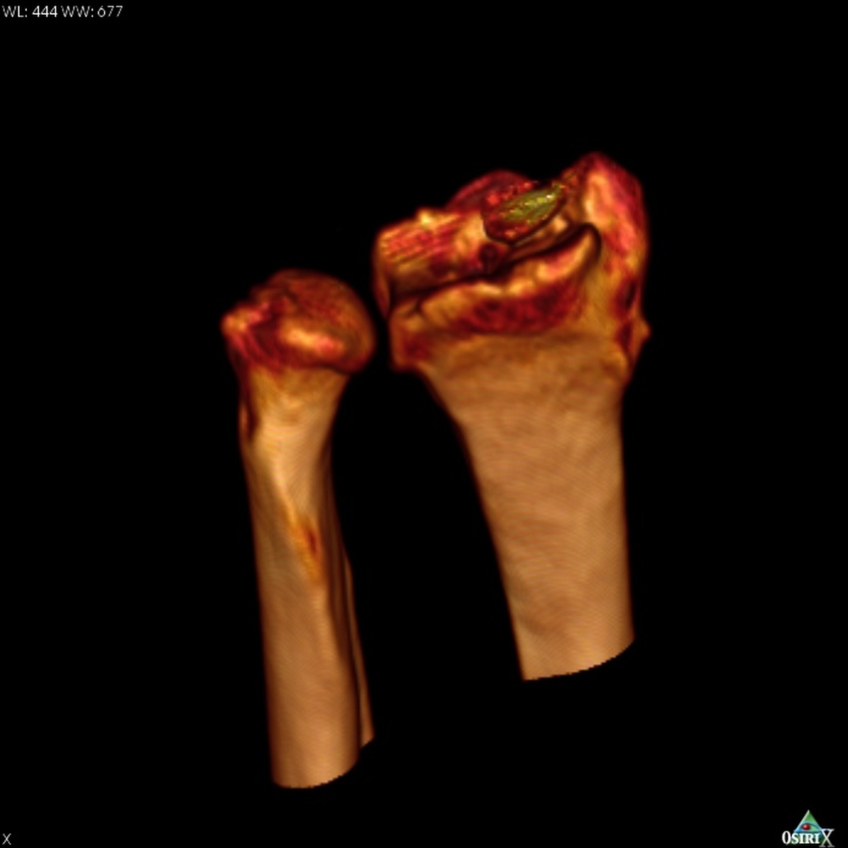
| On first glance, 3D CT
reconstruction looked favorable for possible simple dorsal opening
wedge corrective osteotomy. |
| Below, a fly-around video of the 3D reconstruction: |
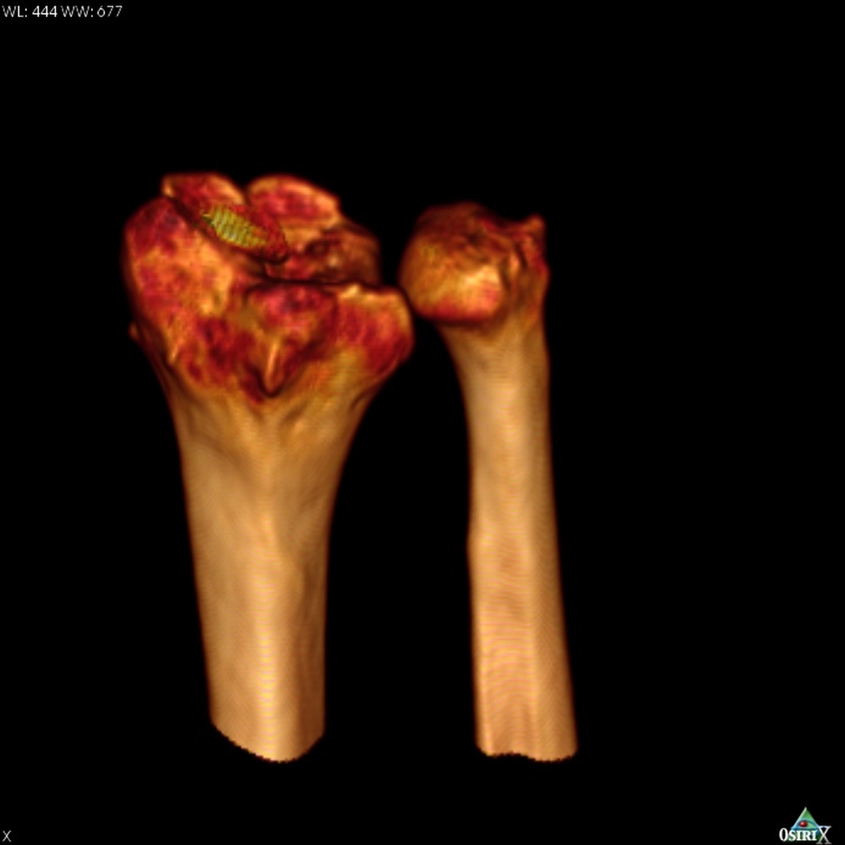
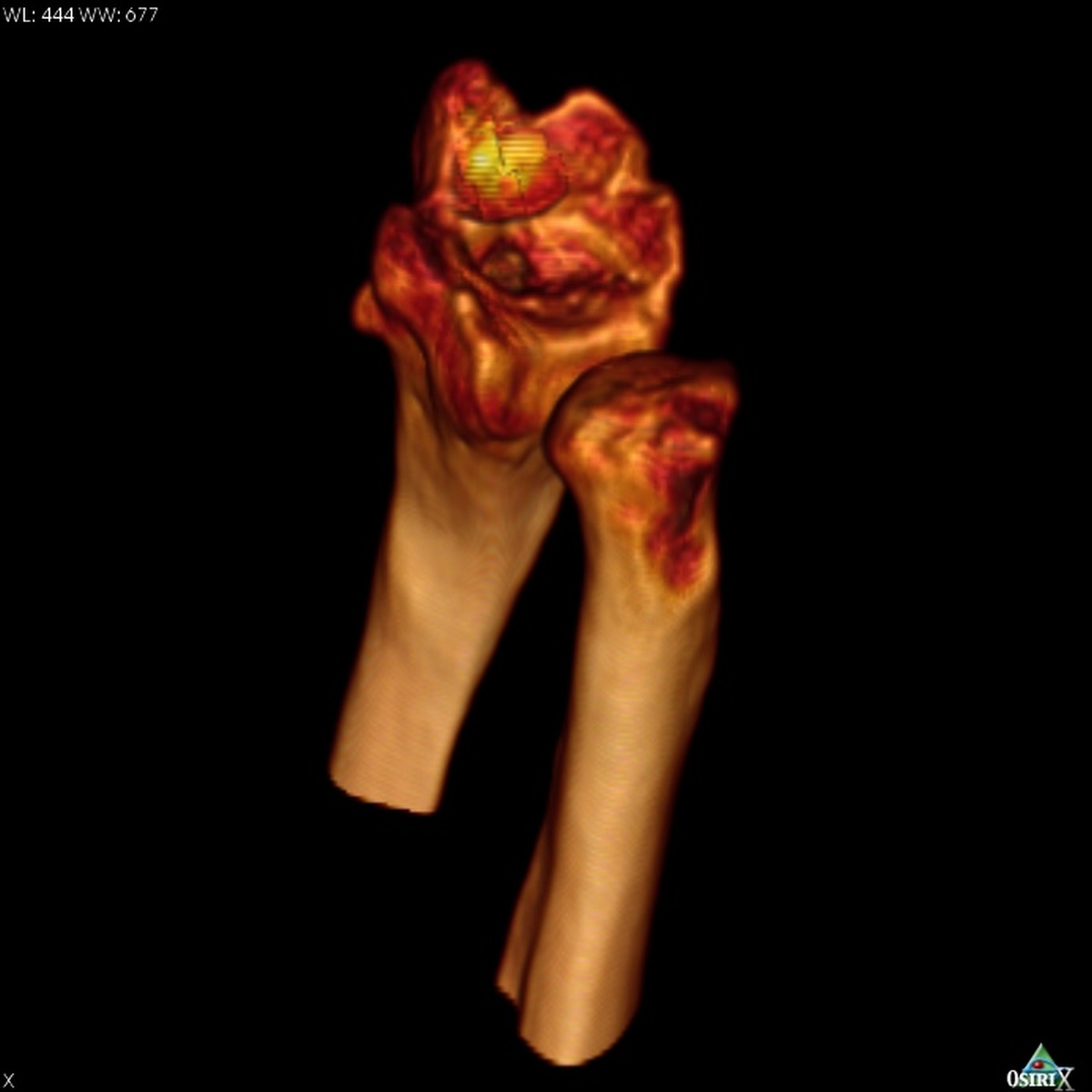
| Digitally hiding the carpal bones obstructing the view of the distal radius shows a much more significant articular defect. In these views, the
proximal cortex of the scaphoid remains as a yellowish shell - it could not be cleanly
removed because it is in bony contact with the dordal lip of the radius. |
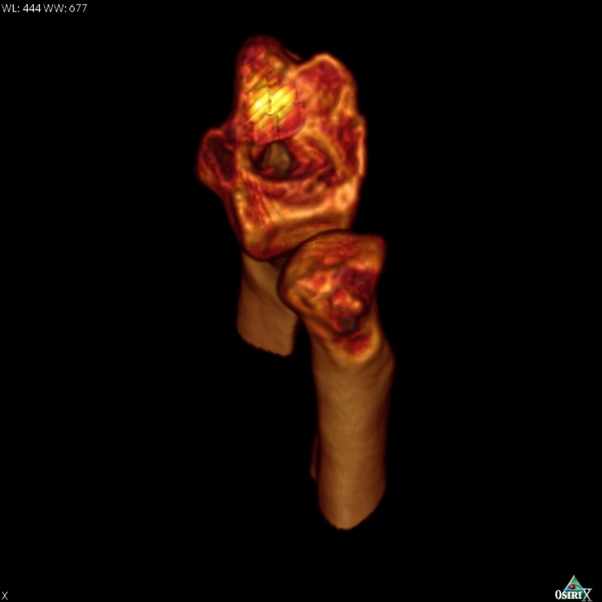
| Video fly around view of the
distal radius articular surface: |
|
Search for... intraarticular distal radius malunion distal radius malunion |
Case Examples Index Page | e-Hand home |