Clinical Example: Knuckle Pads: Dorsal Dupuytren Nodules
| Patients with
Dupuytren's disease sometimes also have localized firm
areas beneath the skin dorsal to the finger joints.
These may or may not be fixed to the skin or to the
underlying extensor mechanism. The overlying skin may be
normal, wrinked or somewhat thin. Such lumps are referred to as ""knuckle pads" or "dorsal Dupuytren nodules" to distinguish them from simple callus or skin thickening over the joints, which are called "dorsal cutaneous pads". Knuckle pads are associated with Dupuytren's and with treatment-resistant Dupuytren's; dorsal cutaneous pads are not. This has been described in more detail by Rayan. Knuckle pads are variable. They:
|
| Click on each image for a larger picture |
| Typical appearing knuckle pads on the middle and ring finger PIP joints of this 38 year old man. He has no evidence of Dupuytren's disease. |
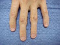
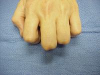
| These are fixed to
the extensor mechanism and are more prominent in PIP
flexion. |
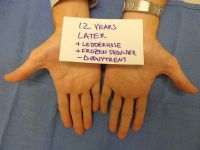
| 12 years later,
the patient has developed Ledderhose disease of one
foot and is recovering from an episode of frozen
shoulder. Both diagnoses are associated with
Dupuytren's disease, but he still has no evidence of
this. |
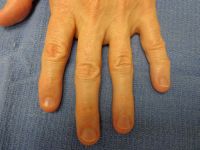
| His initial
knuckle pads have resolved, but he has a new knuckle
pad on the dorsum of his index PIP joint. |
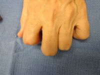
| He may or may not
ever develop palmar Dupuytren's disease. |
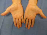
| Woman with
multiple central knuckle pads and Dupuytren's
contracture. |
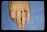
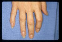
| Another patient
with condylar knuckle pads. |
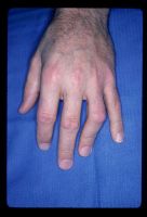
| This patient has
knuckle pads of every PIP joint but no fixed
contractures. |
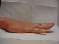
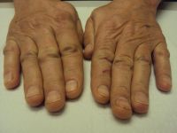
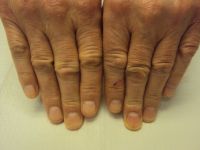
| This patient has
knuckle pads with some dimpling and loss of extension
creases on the small finger. |
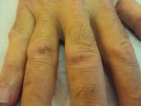
| Knuckle pad of the
DIP joint. |
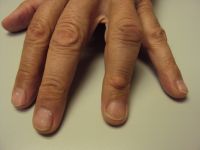
| Tight skin
associated with early aggressive Dupuytren's. |
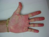
| Ectopic
Dupuytren's is associated with aggressive, recurrent,
treatment resistant Dupuytren's. Knuckle pads are one
ectopic location. less common locations include the
pisiform / distal FCU sheath, as shown here. |
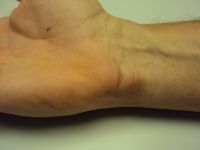
| Knuckle pads on
the same patient, showing some dimpling and skin
flaking. |
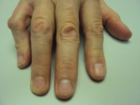
| Another patient
with pisiform area involvement, showing scuff marks
from a recent fall. |
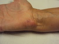
| Same hand
showing inflammatory appearance of knuckle pads with
some loss of skin extensor creases. |
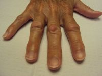
| Black woman with a
new index finger knuckle pad. She had a similar
knuckle pad on the middle finger of the same hand
which resolved without recurrence after an
intralesional steroid injection five years earlier. |
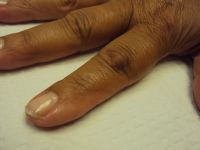
| DIP knuckle pad
resembling a mucous cyst. |
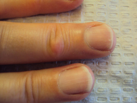
| Another patient
showing crease deformation. |
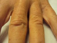
| Same patient,
other hand. |
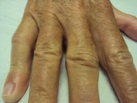
| Atypical very
localized knuckle pad with loss of extensor skin
creases. |
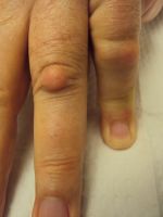
| Same patient,
other hand. |
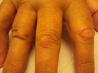
| Another patient,
similar pattern to above. |
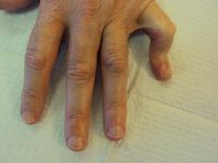
| Well circumscribed knuckle pads with peripheral skin puckering. |
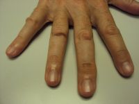
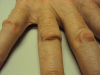
| Circumscribed
knuckle pad with fine dermal creases. |
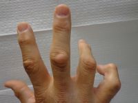
| Unlike a dorsal
cutaneous pad, the epidermis is not hyperkeratotic. |
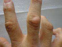
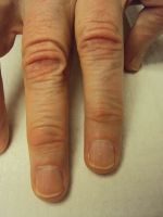
| A variation is deformation or puckering of the skin creases without a mass effect, as in this patient. |
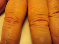
| Same patient,
other hand. |
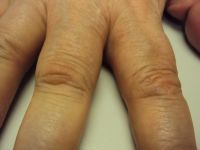
| Another example of
this type of skin puckering. |
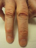
| Same patient other
hand. |
| Central and
lateral knuckle pads, top and side views. |
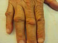
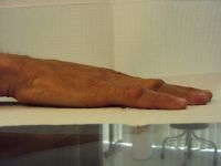
| Three views of a
hand with both single and double knuckle pads and a knuckle pad of the thumb IP joint. |
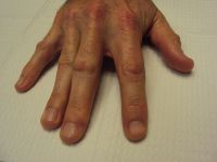
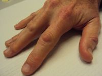
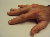
| Knuckle pads with
loss of extensor skin creases. |
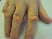
| "Active" knuckle
pads: pink and tender. |
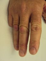
| Knuckle pad
dimpling and prominence. |
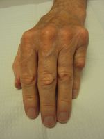
| Bilateral knuckle
pads. |
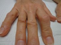
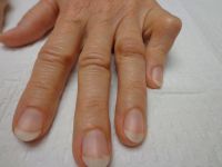
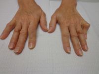
| "Active"knuckle pads, pink and tender, both hands |
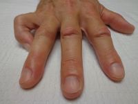
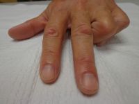
| A patient with
multiple knuckle pads and diffuse palmar involvement
but no contractures - yet. This is the stage at which
a biologic, disease-modifying therapy would be
appropriate, before disabling structural changes
occur. Such a treatment does not exist - yet. |
| Search for... knuckle pads dorsal dupuytren nodules
|
Case Examples Index Page | e-Hand home |